
Abstract
Pulmonary embolism (PE) can rarely mimic an inferior ST-segment elevation myocardial infarction with atrioventricular (AV) block. A 66-year-old woman with morbid obesity collapsed after sudden chest pain and dyspnea. After prehospital and recurrent in-hospital cardiac arrests, she remained intubated and in profound shock. The initial electrocardiogram showed inferior ST elevation, reciprocal changes, and second-degree AV block (Mobitz type 1). Emergency coronary angiography, performed after heparin and cangrelor administration, did not demonstrate an obstructive culprit. Subsequently, bedside echocardiographic reassessment revealed a central echogenic mass at the main pulmonary artery bifurcation despite initially limited signs of right ventricular pressure overload. Computed tomography pulmonary angiography was unsafe because of hemodynamic instability; catheter pulmonary angiography confirmed central obstruction and complete left pulmonary artery occlusion. Mechanical fragmentation using a pigtail catheter was unsuccessful; aspiration was not performed. Following individualized assessment of marked bleeding risk, 20 mg alteplase was administered locally as a non-standard salvage regimen (10 mg into the right pulmonary artery, followed by 10 mg into the left, over approximately 2 minutes per side), without a subsequent infusion. Refractory shock persisted, vascular access site bleeding developed, and the patient subsequently died. Bedside reassessment after culprit-negative coronary angiography identified the cause of refractory shock; the optimal reperfusion approach remains uncertain.
Keywords: Atrioventricular block, case report, echocardiography, pulmonary embolism, ST-segment elevation myocardial infarction
Introduction
Acute pulmonary embolism (PE) can mimic ST-segment elevation myocardial infarction (STEMI), usually with anterior rather than inferior ST elevation (). Inferior STEMI-like PE with culprit-negative coronary angiography has been reported (,), as have PE-associated atrioventricular (AV) conduction disturbances (,). Direct visualization of central pulmonary arterial thrombus by transthoracic echocardiography (TTE) is uncommon (,). Inferior ST elevation with conduction block can be indistinguishable from a coronary emergency during initial resuscitation.
After cardiac arrest, an electrocardiogram (ECG) that appears coronary may prompt urgent angiography before PE is suspected. We describe the diagnostic transition from inferior ST-elevation and AV block to negative coronary angiography, the bedside detection of a central pulmonary arterial mass, the confirmatory catheter angiography, and the difficult reperfusion decisions. The timing of the transition is central to its clinical teaching point.
Case Report
A 66-year-old woman (body mass index of 41 kg/m2) without documented cardiovascular disease, prior thromboembolism, malignancy, or recent surgery collapsed following the abrupt onset of chest pain and dyspnea. Prehospital cardiopulmonary resuscitation (CPR) lasted approximately 15–20 minutes before the return of spontaneous circulation (ROSC; ~21:50). Multiple rib fractures were observed. At the referring hospital (~22:10), blood gases showed pH 6.98, bicarbonate 8.9 mmol/L, lactate 11.8 mmol/L, PaO2 88.2 mmHg, and PaCO2 43.0 mmHg; the ECG showed inferior ST elevation. She was intubated and transferred while receiving vasoactive support, approximately 1 hour after presentation to the referring hospital. A recurrent arrest shortly after arrival required approximately 10 minutes of CPR ().
Table 1. Approximate clinical timeline
Time* | Clinical event and diagnostic/therapeutic action |
|---|---|
Symptom onset | Sudden chest pain, dyspnea, and collapse at home; exact onset time unknown. |
~21:50 | First ROSC after approximately 15-20 min of prehospital CPR. |
~22:10 | Referring-hospital assessment; initial ECG and arterial blood gases. |
~1 h after referring-hospital presentation | Transfer to treating center; recurrent arrest shortly after arrival and ~10 min further CPR. |
~23:30 | Working diagnosis of inferior STEMI; unfractionated heparin and intravenous cangrelor. |
~23:35 | Emergency coronary angiography: no obstructive culprit lesion. |
~23:45-00:00 | Immediate bedside reassessment: central pulmonary arterial mass visualized on TTE; CTPA deferred due to instability. |
~00:30 | Pulmonary angiography in AP and LAO 30° projections: complete left pulmonary artery occlusion. |
After pulmonary angiography | Unsuccessful 5-Fr pigtail mechanical fragmentation; no aspiration performed; informed discussion/consent for local alteplase. |
~00:50 | Local alteplase: right PA 10 mg over ~2 min, then left PA 10 mg over ~2 min (20 mg total); no further infusion. |
~01:30 | Follow-up ECG and TTE: resolved AV block and evolving RV failure. |
~03:30 | Vascular access-site bleeding and hemoglobin decline; O RhD-negative red cells requested but not available before death. |
~06:00 | Death during persistent refractory shock. |
*All clock times are approximate and relative to the presenting episode. CPR: Cardiopulmonary resuscitation, CTPA: Computed tomography pulmonary angiography, ECG: Electrocardiogram, ROSC: Return of spontaneous circulation, STEMI: ST-segment elevation myocardial infarction, TTE: Transthoracic echocardiography
After ROSC, she remained intubated, ventilated, and hypotensive (approximately 70/40-60/30 mmHg) despite an infusion of noradrenaline at 0.06 µg/kg/min. The repeat blood gas showed pH 7.19, bicarbonate 18.1 mmol/L (measured after sodium bicarbonate), lactate 12.58 mmol/L, PaO2 62.2 mmHg, PaCO2 48.6 mmHg, and arterial oxygen saturation 84.4%. The referring hospital ECG showed sinus rhythm with second-degree AV block (Mobitz type 1 [Wenckebach], 4:3 conduction); a ventricular rate of approximately 44 beats/min; inferior ST elevation (II, III, aVF) with reciprocal ST depression in leads I and aVL; and subtle ST elevation in V1 (). The STEMI pathway was activated. The inferior ECG pattern and persistent shock initially made acute coronary occlusion a plausible working diagnosis.
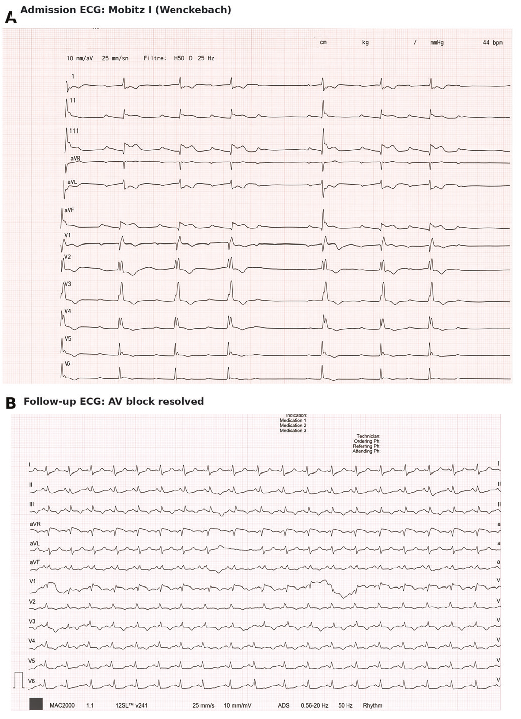
Electrocardiographic evolution. (A) Initial 12-lead ECG obtained at the referring hospital showing second-degree AV block, Mobitz type 1 (Wenckebach), with 4:3 AV conduction and a ventricular rate of approximately 44 beats/min, inferior ST-segment elevation in leads II, III and aVF with reciprocal ST depression in I and aVL, and subtle ST elevation in V1. (B) Follow-up ECG during ongoing reperfusion and hemodynamic management demonstrating sinus tachycardia, resolution of AV block, S1Q3T3, new right bundle branch block, and T-wave inversion in V1-V4
ECG: Electrocardiogram, AV: Atrioventricular
For suspected inferior STEMI, 10,000 IU unfractionated heparin and intravenous cangrelor were administered at ~23:30, before femoral coronary angiography (~23:35), which found no obstructive culprit (, ). The coronary findings did not explain the severity of shock. High-sensitivity troponin I was 904 ng/L (reference <26), BNP was 3000 pg/mL, and D-dimer was 30.9 µg/mL; initial hemoglobin was 13.7 g/dL and platelets were 110×109/L.
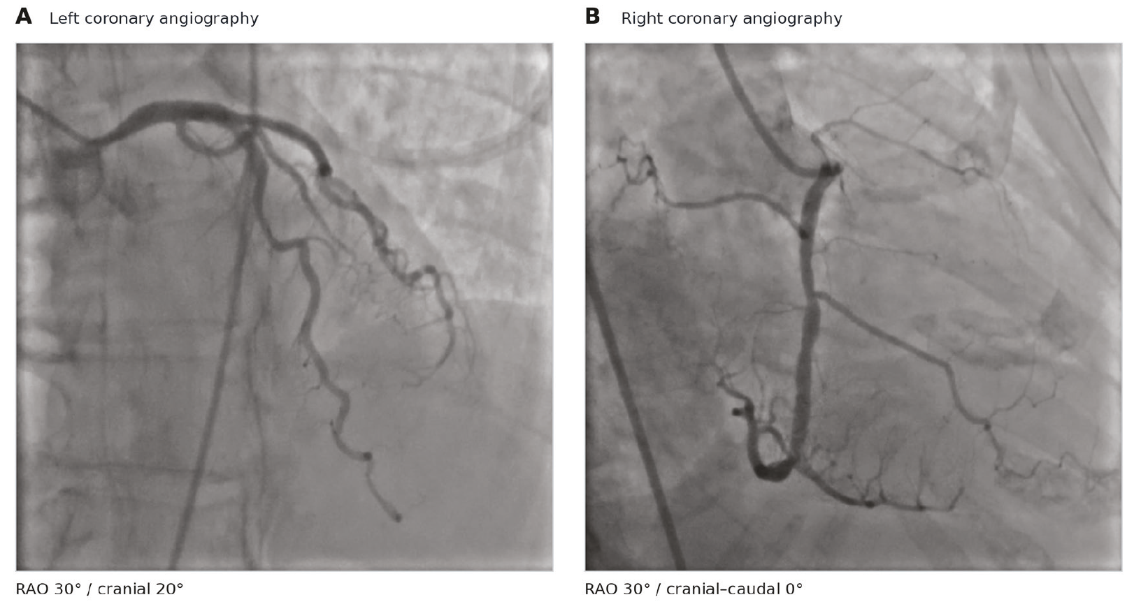
Emergency coronary angiography. (A) Left coronary angiography in a right anterior oblique cranial projection (RAO 30°, cranial 20°) demonstrating no obstructive culprit lesion. (B) Right coronary angiography in a right anterior oblique projection (RAO 30°, cranial/caudal 0°) demonstrating no obstructive culprit lesion. The displayed epicardial vessels are patent without an angiographically identified acute culprit
RAO: Right anterior oblique
Persistent unexplained shock after culprit-negative angiography prompted immediate bedside TTE. Left ventricular systolic function was preserved, without prominent early right ventricular (RV) dilatation or septal flattening. Parasternal short-axis imaging at the great-vessel level revealed a non-mobile echogenic mass (~3.0×1.3 cm) at the main pulmonary artery bifurcation (, ), suggestive of a saddle-like thrombus. PE became the leading diagnosis. Initial tricuspid regurgitation peak velocity was ~2.5 m/s, and the inferior vena cava diameter was 1.3 cm, without visible respiratory collapse. Estimated pulmonary artery systolic pressure was ~25 mmHg, a value considered unreliable during low-flow shock. Computed tomography pulmonary angiography (CTPA) was deferred because transport was unsafe. The bedside mass was considered a trigger for confirmatory PE-directed imaging, but not as definitive proof.
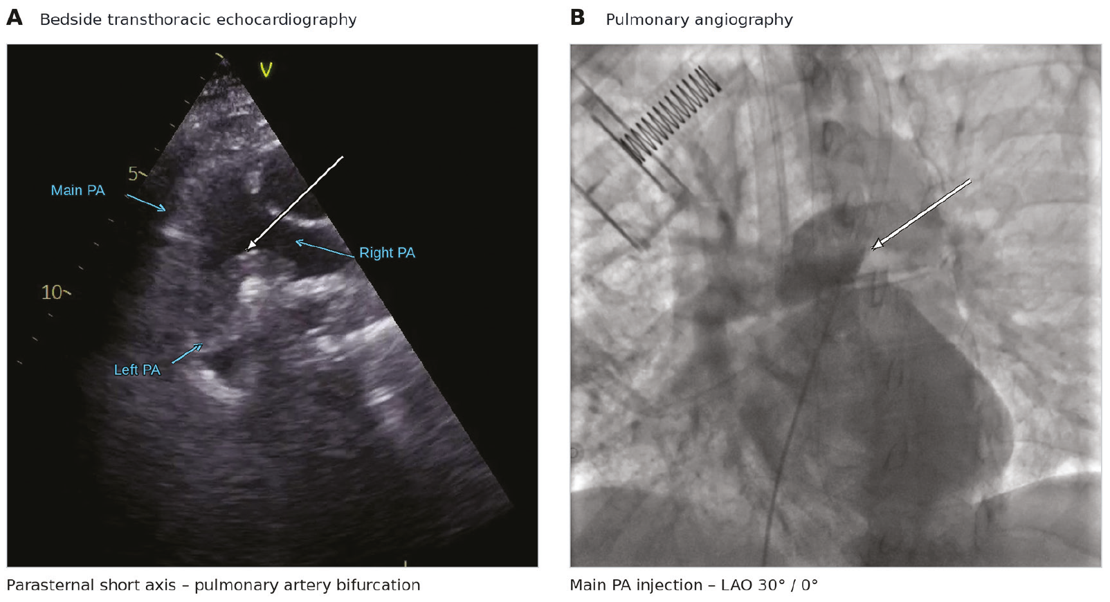
Multimodality identification of central pulmonary embolism. (A) Bedside transthoracic echocardiography in the parasternal short-axis view at the great-vessel level showing a non-mobile echogenic mass (approximately 3.0×1.3 cm) at the main pulmonary artery bifurcation with a saddle-like appearance. The white arrow marks the echogenic mass; blue arrows label the main and branch pulmonary arteries. (B) Catheter pulmonary angiography after contrast injection into the main pulmonary artery, shown in the LAO 30° projection (no cranial/caudal angulation), demonstrating central filling defect and complete left pulmonary artery occlusion; an AP 0°/0° view was also acquired. Angiography did not establish definite bilateral saddle extension
LAO: Left anterior oblique, AP: Anteroposterior, PA: Pulmonary artery
For urgent reperfusion, catheter pulmonary angiography was performed at ~00:30 via femoral venous access with a 5-Fr pigtail catheter into the main pulmonary artery, after a risk discussion and written procedural consent from next of kin. This strategy permitted confirmation and potential treatment without an additional diagnostic transfer. Anteroposterior (AP 0°/0°) and left anterior oblique (LAO 30°/0°) views showed a central bifurcation filling defect, a predominantly left-sided thrombus, and complete occlusion of the left pulmonary artery (). Angiography did not establish extension into both main branches. Despite the saddle-like TTE appearance, a definite anatomical saddle classification was avoided. Recurrent arrest and refractory vasopressor-dependent shock supported high-risk PE (AHA/ACC acute PE category E2) ().
After heparin/cangrelor and before alteplase, INR was 2.38, fibrinogen was 0.64 g/L, and aPTT was unmeasurable because no clot formed; the team knew these results before treatment. Full-dose systemic fibrinolysis was considered but ultimately not selected after individualized assessment of coagulopathy, traumatic CPR with rib fractures, antithrombotic exposure, and femoral arterial/venous access. Surgical embolectomy and extracorporeal membrane oxygenation were unavailable. Mechanical fragmentation using the 5-Fr pigtail failed to achieve adequate reperfusion; the technical reason remained undetermined. No aspiration thrombectomy was performed. The hemorrhagic risks were considered cumulatively; no single risk factor was presented as an absolute contraindication. In the setting of refractory shock, the team discussed the risk of major bleeding and potential benefits with the next of kin, and obtained written consent. At ~00:50, 20 mg of alteplase was delivered locally through the pigtail catheter: 10 mg into the right pulmonary artery over ~2 minutes, followed by 10 mg into the left pulmonary artery over ~2 minutes. No further alteplase bolus or infusion was given. This was a non-standard salvage regimen. The exact distal catheter-tip position during each injection was not separately recorded.
Hypotension worsened despite escalation of noradrenaline from 0.06 to 1.5 µg/kg/min and addition of dopamine (~2.8 µg/kg/min). At ~01:30, AV block had resolved during ongoing management; the ECG showed sinus tachycardia, S1Q3T3, new right bundle branch block, and anterior T-wave inversion (). Repeat TTE showed RV dilatation, septal flattening with a D-shaped left ventricle, significant tricuspid regurgitation, a pulmonary acceleration time of ~61 ms, and an inferior vena cava diameter of 1.9 cm without respiratory collapse (). Estimated pulmonary artery systolic pressure was ~30 mmHg but was unreliable in low-flow RV failure. At ~03:30, oozing was noted from the femoral and intravenous access sites, and local dressings were applied. Hemoglobin decreased from 13.7 g/dL to 10.6 g/dL and then to 6.5 g/dL. Packed red cells were requested; however, O RhD-negative units were unavailable in the hospital blood bank and had not arrived before death at ~06:00. The source of the hemoglobin decline was not fully established, and the contribution of bleeding to death during refractory shock remains uncertain.
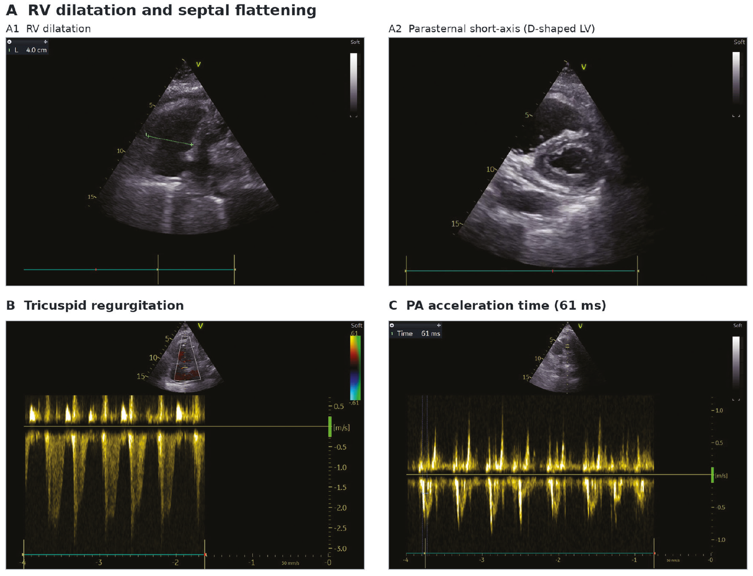
Echocardiographic markers of right ventricular (RV) failure. (A) Repeat transthoracic echocardiography showing RV dilatation (A1) and interventricular septal flattening with a D-shaped left ventricle in the parasternal short-axis view (A2). (B) Doppler echocardiography demonstrating tricuspid regurgitation. (C) Pulmonary artery Doppler showing a shortened acceleration time of approximately 61 ms. The estimated pulmonary artery systolic pressure (~30 mmHg) was interpreted cautiously in the setting of severe low-flow RV failure
PA: Pulmonary artery
Discussion
Inferior STEMI-like PE with culprit-negative coronary angiography (,), PE-associated AV block (,), and central thrombus detected by TTE (,) have each been reported. Our case emphasizes their diagnostic sequence after arrest: an apparent coronary event, negative angiography, an immediate, focused TTE, and confirmatory pulmonary angiography. Direct thrombus visualization was especially useful because conventional RV strain findings were initially limited.
RV pressure overload or ischemia, supply-demand mismatch, hypoxemia, and post-arrest ischemia may contribute to ST changes (,,). A transient coronary event or vasospasm cannot be excluded on the basis of a later culprit-negative angiogram. AV block may reflect RV ischemia, autonomic effects, or post-arrest physiology; its resolution during treatment does not establish a causal effect of alteplase. This was a STEMI-like presentation with confirmed PE; it does not prove a single mechanism. The serial ECGs document the evolution but cannot identify a unique electrophysiological pathway.
Echocardiography alone cannot confirm or exclude PE because indirect RV findings lack specificity (). Here, direct bifurcation-mass visualization, despite limited early RV signs, prompted catheter angiography. The saddle-like TTE appearance was not definitive; angiography confirmed central obstruction and left pulmonary artery occlusion but did not show unequivocal bilateral branch extension. Similar bedside ultrasound redirection has been described (). Angiography confirmed the obstruction but did not resolve all anatomical uncertainties suggested by TTE.
Advanced reperfusion in PE with cardiopulmonary failure depends on the hemorrhagic risk and on local resources (,). Known pre-alteplase coagulation abnormalities, traumatic CPR, antithrombotic exposure, and femoral access increased bleeding risk, but did not individually constitute absolute contraindications to systemic thrombolysis. With surgery and extracorporeal support unavailable, and fragmentation unsuccessful, the team selected rapid local administration of alteplase after a documented risk-benefit discussion and written consent. The total alteplase dose (20 mg) was administered over approximately 4 minutes without further infusion; however, this is not an established catheter-directed thrombolysis protocol. Subsequent bleeding had multiple contributing factors and no single identifiable cause. The effect of this regimen on survival or bleeding cannot be determined from a single case.
Limitations include unavailability of CTPA, invasive pulmonary pressure measurements, and autopsy. RV/LV ratio, TAPSE, and RV S′ could not be reliably retrieved from emergency images; therefore, RV enlargement was assessed qualitatively. Access-site oozing did not account for the full extent of the hemoglobin decline; transfusion was not begun before death. Neither the independent contribution of hemorrhage nor the question of whether earlier TTE or a different reperfusion strategy would have changed survival can be determined from a single case.
Conclusion
High-risk PE may mimic inferior STEMI with AV block. After culprit-negative coronary angiography in unstable post-arrest shock, bedside TTE may reveal central PE and guide confirmatory imaging when CTPA is unsafe. TTE cannot independently confirm PE or determine a preferred reperfusion strategy.
Ethics
Informed Consent: Written informed consent for publication of this case report and accompanying electrocardiogram, echocardiographic, coronary angiographic, pulmonary angiographic, and video material was obtained from the patient’s next of kin. The treating team discussed the substantial risks and potential benefits of pulmonary embolism-directed invasive treatment and local thrombolysis with the patient’s next of kin. Written consent was obtained before treatment.
Footnotes
Authorship Contributions
Surgical and Medical Practices - Concept - Design - Data Collection or Processing - Analysis or Interpretation - Literature Search - Writing: All authors contributed equally to this study.
Conflict of Interest
No conflict of interest was declared by the authors.
Financial Disclosure
The authors declared that this study received no financial support.
Acknowledgements
The authors thank Samet Solmaz, MD, Clinic of Cardiology, Kocaeli City Hospital, for assistance with the clinical care of the patient.
Reporting Guideline: This report was prepared in accordance with CARE recommendations; a completed CARE checklist is supplied separately. The patient’s perspective was unavailable because she remained intubated after the arrest and died during the index admission.
Declaration Regarding the Use of AI and AI-Assisted Technologies: During manuscript preparation, ChatGPT (OpenAI) was used for language editing, journal-specific restructuring, and editorial critique. The authors reviewed and verified the final text and take full responsibility for its content.
References
Villablanca PA, Vlismas PP, Aleksandrovich T, Omondi A, Gupta T, Briceno DF, et al. Case report and systematic review of pulmonary embolism mimicking ST-elevation myocardial infarction. Vascular. 2019;27:90-7.
Emren SV, Arikan ME, Senoz O, Varis E, Akan E. Acute pulmonary embolism mimicking inferior myocardial infarction. Turk Kardiyol Dern Ars. 2014;42:290-3.
Mistry A, Natarajan N, Hussain S, Vali Z. Unusual presentation of acute pulmonary embolus presenting with inferior ST elevation. BMJ Case Rep. 2018;2018:bcr2018226063.
Akinboboye OO, Brown EJ Jr, Queirroz R, Cusi VP, Horowitz LF, Jonas EA, et al. Recurrent pulmonary embolism with second-degree atrioventricular block and near syncope. Am Heart J. 1993;126:731-2.
Ma M, Liang S, He Y, Wang H. Case report: The presence of third-degree atrioventricular block caused by pulmonary embolism masquerading as acute ST-segment elevation myocardial infarction. Front Cardiovasc Med. 2023;10:1013727.
Yuriditsky E, Horowitz JM, Taslakian B, Saric M. Saddle pulmonary embolism detected by transthoracic echocardiography in a patient with suspected myocardial infarction. CASE (Phila). 2024;8:54-7.
Lipa AJ, Domanovits H, Schwameis M. Saddle pulmonary embolism detected by transthoracic echocardiography-a case report. Wien Klin Wochenschr. 2026.
Writing Committee Members; Creager MA, Barnes GD, Giri J, Mukherjee D, Jones WS, Burnett AE, et al. 2026 AHA/ACC/ACCP/ACEP/CHEST/SCAI/SHM/SIR/SVM/SVN Guideline for the evaluation and management of acute pulmonary embolism in adults: a report of the American College of Cardiology/American Heart Association Joint Committee on Clinical Practice Guidelines. Circulation. 2026;153:e977-1051.
Pruszczyk P, Klok FA, Kucher N, Roik M, Meneveau N, Sharp ASP, et al. Percutaneous treatment options for acute pulmonary embolism: a clinical consensus statement by the ESC Working Group on pulmonary circulation and right ventricular function and the European Association of Percutaneous Cardiovascular Interventions. EuroIntervention. 2022;18:e623-38.
Shy BD, Gutierrez A, Strayer RJ. Bedside ultrasound to evaluate pulmonary embolism masquerading as ST elevation myocardial infarction (STEMI). J Emerg Med. 2015;49:703-4.e1.
Supplementary Materials
Supplement 1. Supplementary Video 1. Parasternal short-axis cine at the level of the great vessels demonstrating an echogenic mass at the main pulmonary artery bifurcation https://youtu.be/HZ05crcval4
Download